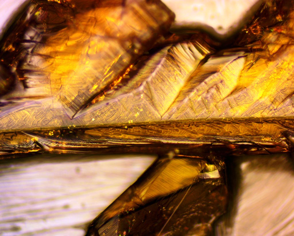
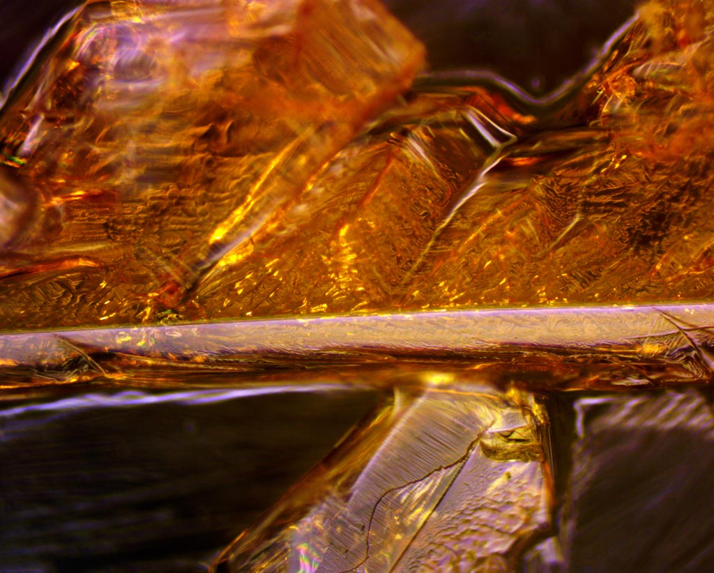
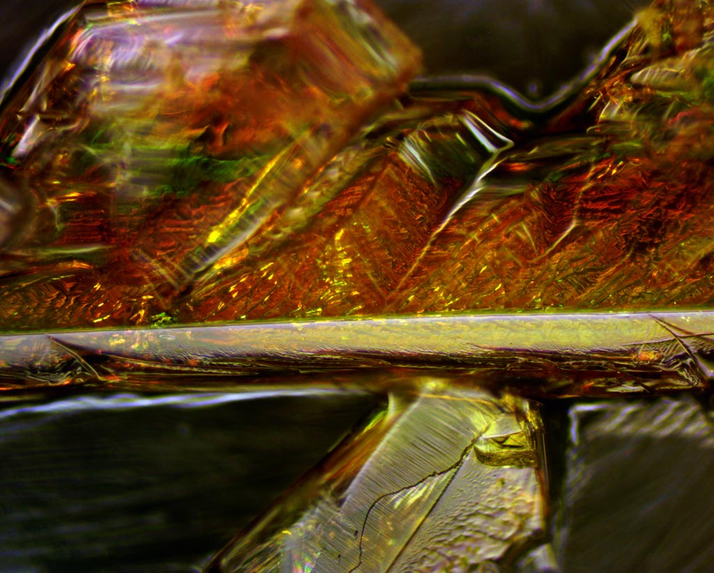
The above images show simple brightfield and darkfield episcopic views of the potassium ferricyanide crystals. It is seen that the crystals are yellowish. While the light source is certainly episcopic (the objective is also acting as the condenser), it is not exactly "reflected", as what we are seeing is light that is penetrating through the crystals and being reflected / scattered from the bottom surface and the crystal itself. As we will see below, they are highly birefringent, so are an interesting subject for polarized light microscopy.
For the polarization comparison, we start with parallel and crossed polarizers, with the analyzer fixed at about 45° with respect to the x-y axes of the image, and the input linear polarizer able to be rotated. Some light is lost but many features of parallel polarizers are very similar to the image with brightfield illumination. Note however, that some colors are visible where the plane of polarization is rotated or becomes elliptical. With crossed polarizers, similar, but effects occur, but with better contrast - note that red and green regions are interchanged.
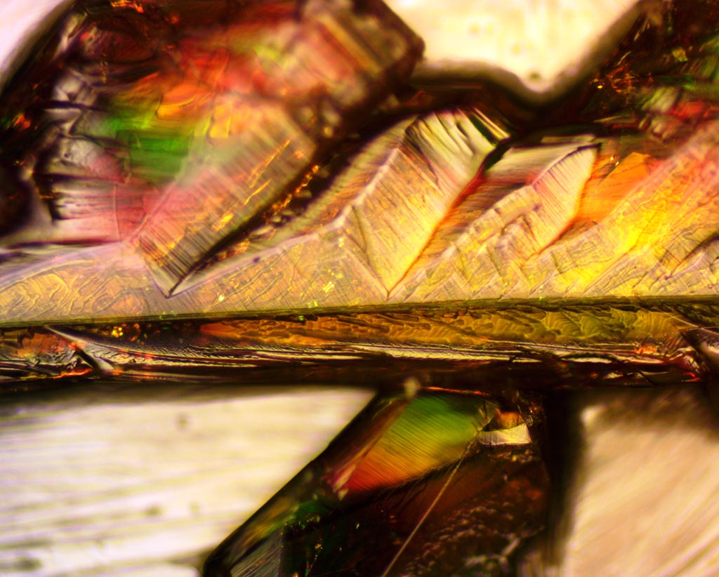
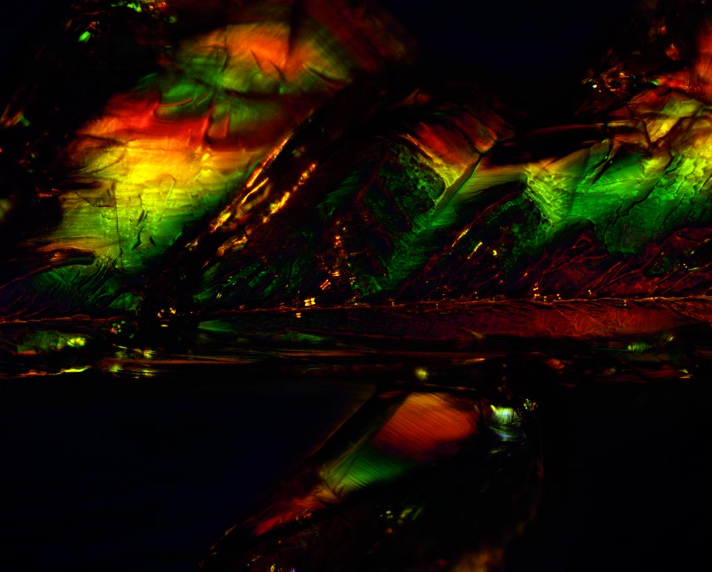
The input polarizer rotation can be seen in this animated .gif (6.9 MB .gif) with frames every 10°.
Next, we have the same comparison, but through the darkfield part of the BD Plan objective.
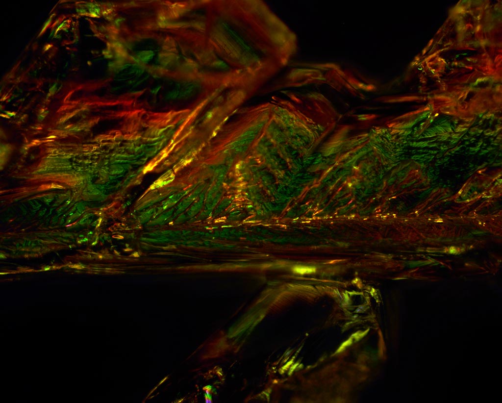
The input polarizer rotation can be seen in this animated .gif (8.2 MB .gif) with frames every 10°.
Now, we see what the darkfield looks like at a single wavelength, through a green interference filter (GIF). Looking at several monochromatic images, one could detangle the multiple-order retardance to determine the actual values of retardance.
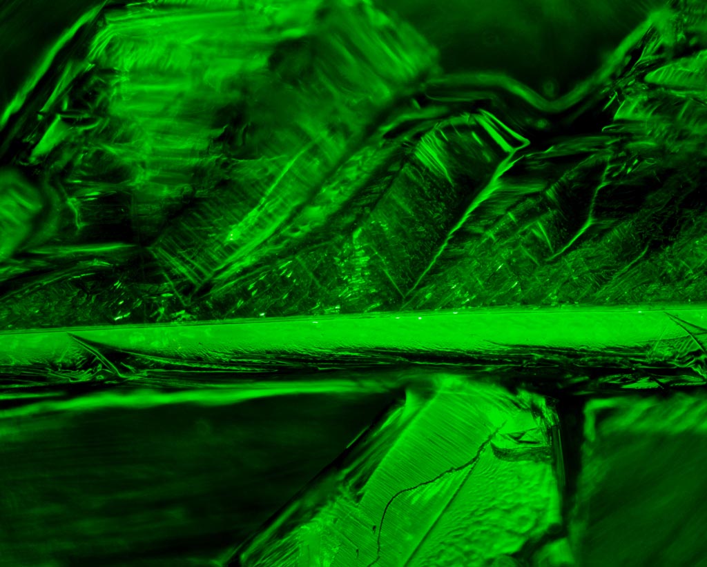
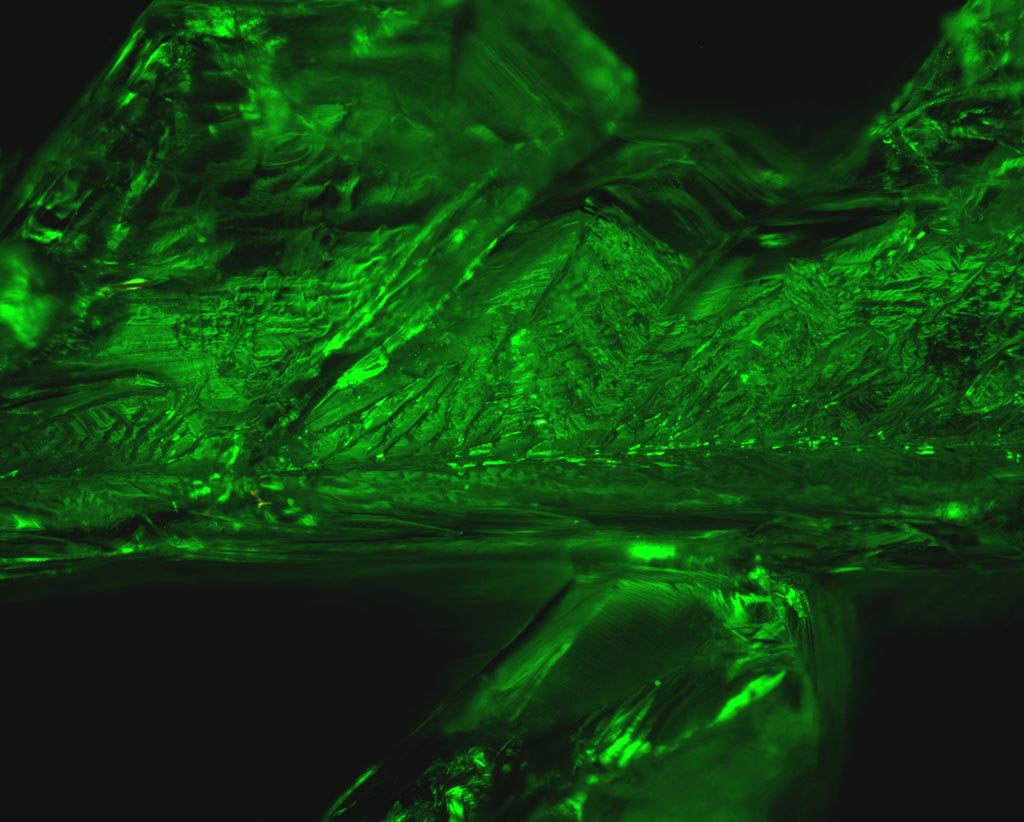
The input polarizer rotation can be seen in this animated .gif (7.1 MB .gif) with frames every 10°.
Thus far, much of the crystal axis has been aligned horizontally, being 45° or so from the angle of the polarizers, which is why colors due to retardance has been displayed. Now I will show the effect of rotation of the specimen (equivalent to rotating the polarizer and analyzer together). First we have the brightfield mode, with the axis generally at 45°, then at 0°. These final images are all with the two polarizers crossed.
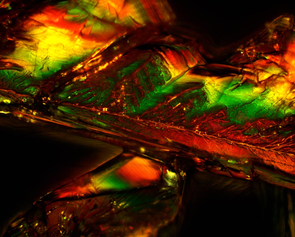
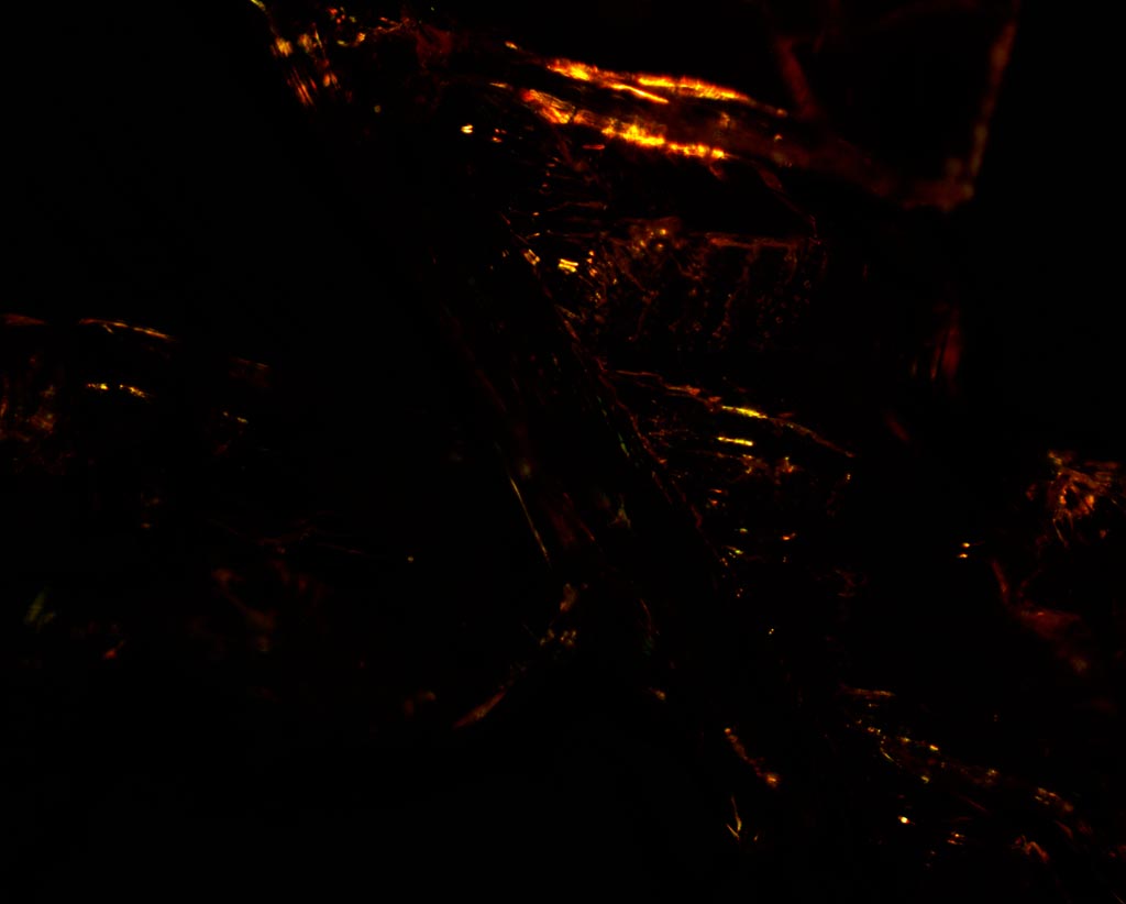
The specimen rotation can be seen (after de-rotation) in this animated GIF (6.7 MB .gif) with frames every 15°.
Here we see the same thing, except in darkfield.
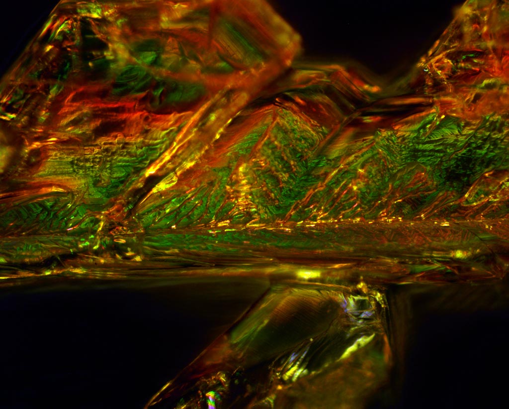
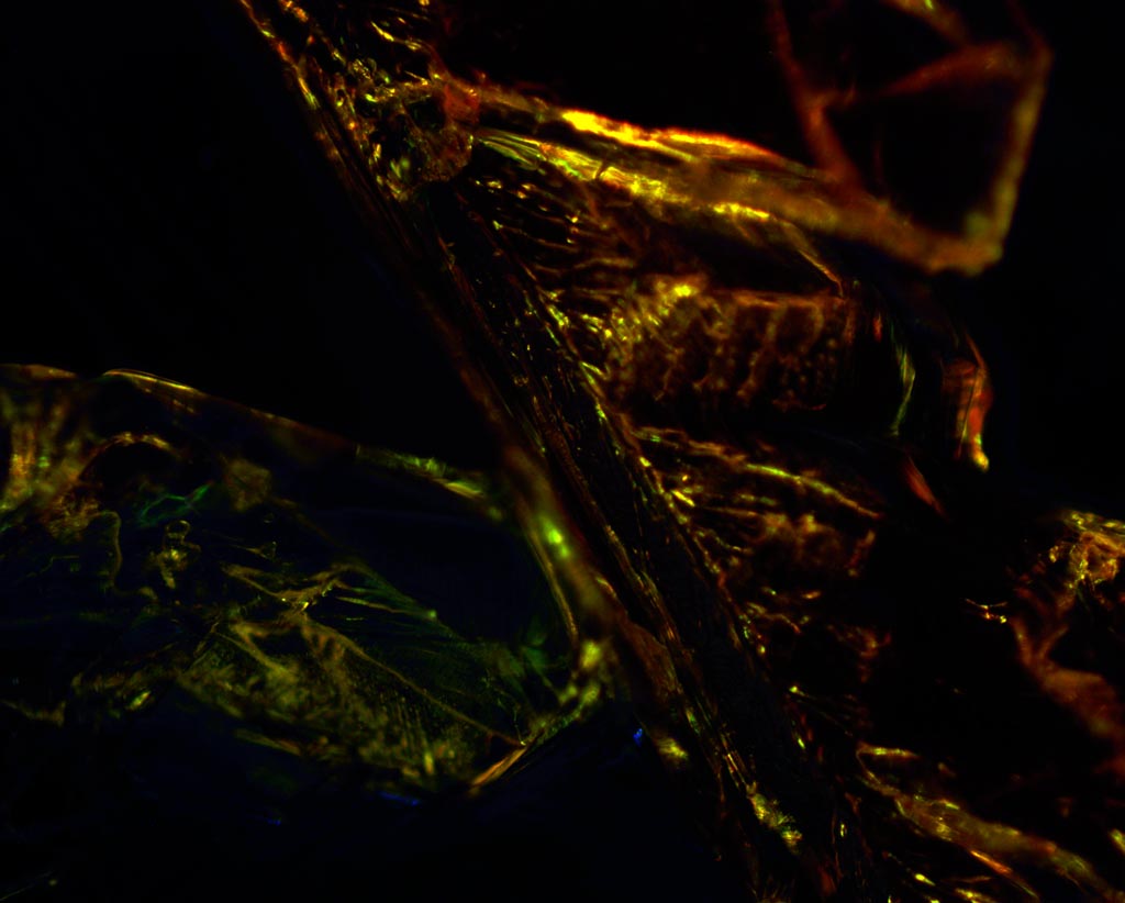
The specimen rotation can be seen (after de-rotation) in this animated GIF (7.9 MB .gif) with frames every 15°.
In the final two comparisons, we can see that with crossed polarizers, certain specimen rotation angles result in almost no light getting back into the objective, meaning that the polarization axis is aligned with the crystal fast (or slow) axis, which means that the polarization state is unchanged, so is blocked by the analyzer. Darkfield has more structure visible because of the high NA scattering of the darkfield illumination.